My appointment is on Tuesday morning, 2018.07.17. I need to arrive by 9:00 a.m. to be prepared for the actual 3-D Imaging at 10:00 a.m. The CCTA Imaging consists of the following overview:

| CCTA 3D Imaging of the Heart ~ 2018.07.17 | ||
| Space. | ||
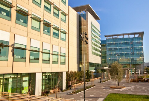
UCSF Hospital - Misson Bay - 1975 4th Street - C1422 |
||

|
On June 11th, 2018, I was called by the General Hospital in conjunction with the VA Hospital to see if I could sign onto various research projects.
I met Danny, Dr. Pricilla Hsue, Brendan Neilan and Smruti Rahalkar as they went over the forms and what research projects were in the works.
This is the first of numerous research projects I will be doing for the UCSF Medical Center -Misson Bay. My contact at San Francisco General Hospital is Smruti Rahalkar, MAS, as a Senior CRC - Dept. of Medicine at 1001 Potrero Avenue 5G4 - San Francisco, CA - Fax: 1.415.206.5461
My appointment is on Tuesday morning, 2018.07.17. I need to arrive by 9:00 a.m. to be prepared for the actual 3-D Imaging at 10:00 a.m. The CCTA Imaging consists of the following overview: |

|
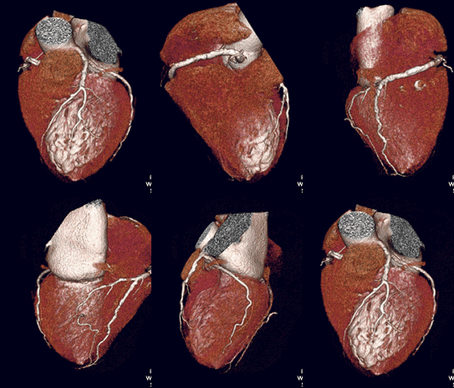
Coronary Computed Tomography Angiography (CCTA)Coronary computed tomography angiography (CCTA) uses an injection of iodine-rich contrast material and CT scanning to examine the arteries that supply blood to the heart and determine whether they have been narrowed by plaque buildup. The images generated during a CT scan can be reformatted to create three-dimensional images that may be viewed on a monitor, printed on film, or transferred to electronic media.

Tell your doctor if there’s a possibility you are pregnant and discuss any recent illnesses, medical conditions, medications you’re taking, and allergies. You will be instructed not to eat or drink anything several hours beforehand and to avoid caffeinated products, Viagra or similar medication. If you have a known allergy to contrast material, your doctor may prescribe medications to reduce the risk of an allergic reaction. These medications must be taken 12 hours prior to your exam. Leave jewelry at home and wear loose, comfortable clothing. You may be asked to wear a gown. If you are breastfeeding, talk to your doctor about how to proceed.

Coronary computed tomography angiography (CCTA) is a heart imaging test that helps determine if plaque buildup has narrowed a patient's coronary arteries, the blood vessels that supply the heart. Plaque is made of various substances circulating in the blood, such as fat, cholesterol and calcium that deposit along the inner lining of the arteries. Plaque, which builds up over time, can reduce or in some cases completely block blood flow. Patients undergoing a CCTA scan receive an iodine-containing contrast material (dye) as an intravenous (IV) injection to ensure the best possible images of the heart blood vessels.
Computed tomography, more commonly known as a CT or CAT scan, is a diagnostic medical test that, like traditional x-rays, produces multiple images or pictures of the inside of the body.
The cross-sectional images generated during a CT scan can be reformatted in multiple planes, and can even generate three-dimensional images. These images can be viewed on a computer monitor, printed on film or transferred to a CD or DVD.
CT images of internal organs, bones, soft tissue and blood vessels provide greater detail than traditional x-rays, particularly of soft tissues and blood vessels.
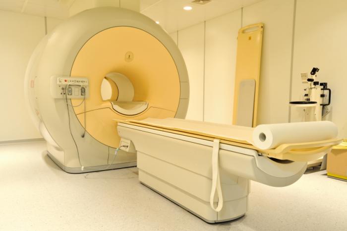
What does the equipment look like?The CT scanner is typically a large, box-like machine with a hole, or short tunnel, in the center. You will lie on a narrow examination table that slides into and out of this tunnel. Rotating around you, the x-ray tube and electronic x-ray detectors are located opposite each other in a ring, called a gantry. The computer workstation that processes the imaging information is located in a separate control room, where the technologist operates the scanner and monitors your examination in direct visual contact and usually with the ability to hear and talk to you with the use of a speaker and microphone.
CCTA is very much like a normal CT scan. The only difference is the speed of the CT scanner and the use of a heart monitor to determine your heart rate.
How does the procedure work?During the examination, x-rays pass through the body and are picked up by special detectors in the scanner. Typically, higher numbers (especially 64 or more) of these detectors result in clearer final images. For that reason, CCTA often is referred to as multi-detector or multi-slice CT scanning.
The information collected during the CCTA examination is used to identify the coronary artery anatomy, plaque, narrowing of the vessel, and, in certain cases, heart function.
The radiologist will use the computer to create three-dimensional images and images in various planes to completely evaluate the heart and coronary arteries.
When a contrast material is introduced to the bloodstream during the procedure, it clearly defines the blood vessels being examined by making them appear bright white.
Space.
Created on: 2018.06.11

Updated on: 2018.07.17