I was asked by the VA Hospital
if I wanted to have an ultra-sound taken of the blood vessels in my neck
to determine if I might have plaque. I agreed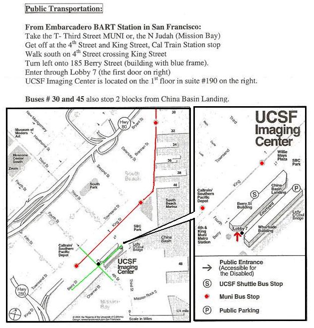 and on 12.14.2007, I
 had the ultra-sound at General Hospital. It was very interesting, they had
me lie down and then proceeded to put the gel on my neck and then did the
left side first, then the right side. I was able to see them doing
the imaging and one could see the blood vessel walls and also when imaged
in color, you could see the red blood going into the head and cooler blue
blood coming back to the heart. You could also hear the pumping of
the blood through the vessels! My heart rate after I was lying down
was at 61. I was relaxed. They also did a short imaging of my
heart just in case I qualified for further testing.
had the ultra-sound at General Hospital. It was very interesting, they had
me lie down and then proceeded to put the gel on my neck and then did the
left side first, then the right side. I was able to see them doing
the imaging and one could see the blood vessel walls and also when imaged
in color, you could see the red blood going into the head and cooler blue
blood coming back to the heart. You could also hear the pumping of
the blood through the vessels! My heart rate after I was lying down
was at 61. I was relaxed. They also did a short imaging of my
heart just in case I qualified for further testing.
They asked me if I might want to have my a CT Scan of my heart imaged at the University of California - San Francisco. I agreed and am waiting to hear if I qualify! This would be two separate imaging and I was surprised as I found out I was going to be paid $75 for the first research project and probably will be paid for the MRI also. (This is the new billion dollar bio-tech research center.)
Rebecca Hoh
Clinical Research Manager - UCSF/SFGH
995 Potrero Avenue, Ward 84
San Francisco, CA 94110
VM: 415.476.4082 ext. 139
FAX: 415.476.6953
E-mail: rhoh@php.ucsf.edu
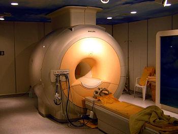
Note: So I went at the appointed hour and I was through in only ten minutes. Interesting, the CT Scan is like an X-Ray. I had to get completely undressed and wear multiple gowns and socks. Then laid on the table and slid into the scanner. A digitized voice said to lie still. Then the machine hummed and stopped. The voice indicated I should take a deep breath and hold it. The machine heads whirled around and the bed moved about two inches at a time. Then I was told to breath again. This happened several times during the 10 minutes in the scanner. I was told I would be informed if there were any areas of plaque that needed taken care of. So far so good! The technician said I probably would be asked to do a second one in the near future.... for another $75. Such a deal!
Heart Disease: Diagnosing Heart Disease: Cardiac Computed Tomography (CT)
Computed
tomography, commonly known as a CT scan, combines multiple X-ray images
with the aid of a computer to produce cross-sectional views of the body.
Cardiac CT is a heart-imaging test that uses CT technology with or without
intravenous (IV) contrast (dye) to visualize the heart anatomy, coronary
circulation, and great vessels (which includes t
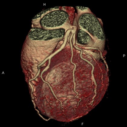 he aorta, pulmonary veins,
and arteries).
he aorta, pulmonary veins,
and arteries).
There are several types of CT scans used in the diagnosis of heart disease such as the Calcium-score screening heart scan that I had.
Calcium-score screening heart scan:
The calcium-score screening heart scan is a test used to detect calcium deposits found in atherosclerotic plaque in the coronary arteries. State-of-the-art computerized tomography methods, such as this one, are the most effective way to detect early coronary calcification from atherosclerosis, before symptoms develop. The amount of coronary calcium has been recognized as a powerful independent predictor of future cardiac events and may be used to guide lifestyle modifications and preventive medical therapies to reduce this risk.
Your doctor uses the calcium-score screening heart scan to evaluate risk for future coronary artery disease. If calcium is present, the computer will create a calcium "score" that estimates the extent of coronary artery disease based on the number and density of calcified coronary plaques in the coronary arteries.
Absence of calcium is considered a "negative" exam. However, since there are certain forms of coronary disease, such as "soft plaque" atherosclerosis, that escape detection during this CT scan, it is important to remember that a negative test indicates a low risk but does not absolutely exclude the possibility of a future cardiac event, such as a heart attack.
The calcium-score screening heart scan takes only a few minutes to perform and does not require injection of intravenous iodine contrast.
