So I went in and was told the test was about 50 minutes long and I couldn't (or shouldn't) move. Into the machine I went, all the way! I guess many get claustrophobia and need to come out. It that happens, one either must do the entire test again or cop out of the test. The machine has huge magnets and the technician told me it consumed about 450 thousands volts of electricity! The voltage builds up and suddenly there is a huge band and the magnets are energized! I was told not to move. I was able to listen to symphonic music as I laid there. I also had a plastic mask over my head, a strange feeling I must say! After the hour I was able to sit down with the technician and view my entire brain, flying through it from one ear to the other. From the side and from the top, through each layer of the brain. Talk about interesting!
MRI
Magnetic Resonance Imaging (MRI) uses
magnetic fields and radio waves to produce high quality two- or
three-dimensional images of brain
 structures without use of ionizing
 radiation (X-rays) or radioactive tracers. During an MRI, a large
cylindrical magnet creates a magnetic field around the head of the patient through which radio waves are sent. When the
magnetic field is imposed, each point in space has a unique radio
frequency at which the signal is received and transmitted (Preuss).
Sensors read the frequencies and a computer uses the information to
construct an image. The detection mechanisms are so precise that changes
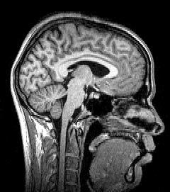
in structures over time can be detected. Using MRI, scientists can create
images of both surface and subsurface structures with a high degree of
anatomical detail. MRI scans can produce cross sectional images in any
direction from top to bottom, side to side, or front to back. The problem
with original MRI technology was that while it provides a detailed
assessment of the physical appearance, water content, and many kinds of
subtle derangements of structure of the brain (such as inflammation or
bleeding), it fails to provide information about the metabolism of the
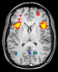
brain (i.e. how actively it is functioning) at the time of imaging. A
distinction is therefore made between "MRI imaging" and
"functional MRI imaging, where MRI provides only
structural information on the brain while MRI yields both structural and
functional data.
radiation (X-rays) or radioactive tracers. During an MRI, a large
cylindrical magnet creates a magnetic field around the head of the patient through which radio waves are sent. When the
magnetic field is imposed, each point in space has a unique radio
frequency at which the signal is received and transmitted (Preuss).
Sensors read the frequencies and a computer uses the information to
construct an image. The detection mechanisms are so precise that changes
in structures over time can be detected. Using MRI, scientists can create
images of both surface and subsurface structures with a high degree of
anatomical detail. MRI scans can produce cross sectional images in any
direction from top to bottom, side to side, or front to back. The problem
with original MRI technology was that while it provides a detailed
assessment of the physical appearance, water content, and many kinds of
subtle derangements of structure of the brain (such as inflammation or
bleeding), it fails to provide information about the metabolism of the
brain (i.e. how actively it is functioning) at the time of imaging. A
distinction is therefore made between "MRI imaging" and
"functional MRI imaging, where MRI provides only
structural information on the brain while MRI yields both structural and
functional data.
